|
Overview
Retrocalcaneal bursitis is the painful inflammation and swelling of the retrocalcaneal bursa that is situated between the calcaneus (heel bone) and the Achilles tendon. A bursa is a small fluid filled sac that forms around joints in areas where there is a lot of friction between muscles, tendons and outcrops of bone. The bursae position themselves in between the tendon or muscle and the bone, buffering any friction from movement. To picture a bursa imagine it as a very small water filled balloon that sits in places where things rub against each other, such as in between a tendon and a bone, to provide a soft smooth cushion for the tendon to pass over painlessly. The covering of the bursa also acts as a lubricant and aids the tendon?s movement. It is estimated that there is over 150 bursae in your body which protect the joint and tendons from wear. They are all very small and unnoticeable until they become swollen and painful with bursitis. Causes The calcaneal bursa can become inflamed in patients with heel spurs or in patients with poor-fitting shoes (eg, high heels). Inflammation can occur secondarily from Achilles tendinitis, especially in young athletes. Patients exhibit tenderness to palpation of the bursa anterior to the Achilles tendon on both the medial and lateral aspects. They have pain with movement, which is worsened with dorsiflexion. Symptoms Pain and tenderness usually develop slowly over time. Applying pressure to the back of the heel can cause pain. Wearing shoes may become uncomfortable. The back of the heel may feel achy. Pain is exacerbated when the foot is pointed or flexed, because the swollen bursa can get squeezed. A person with retrocalcaneal bursitis may feel pain when standing on their toes. Fever or chills in addition to other bursitis symptoms can be a sign of septic bursitis. Though uncommon, septic retrocalcaneal bursitis is a serious condition, and patients should seek medical care to ensure the infection does not spread. Diagnosis A physical examination will be performed to determine if you have any signs of Achilles Bursitis or other ankle injury. He/she will look and feel the soft tissue and bones in your ankles to note any differences between the two of them. This will identify any abnormalities, such as swelling, bone deformities, atrophied muscles, redness and/or warmth on the skin. In many cases, the first sign that you have Achilles bursitis is swelling in the back of the foot and ankle pain. Non Surgical Treatment Most patients with achilles and retrocalcaneal bursitis heal well with appropriate physiotherapy and other usual bursitis conventional and natural cures being administered. Specific treatments for ankle / heel bursitis may include footwear correction. Get well-fitting, soft-backed (or even open-backed whenever possible) shoes for both day to day wear and exercise. High-heels should really be a no no or worn sparingly, ladies. Heel protectors. Heel pads and heel lifts are great simple solutions to cushion and protect the Achilles area from the irritation of the shoes. Orthotics. There are various orthotic devices out there (some only available over-the-counter). One example is a custom arch suppport. These can control abnormal motion in your feet by lining them up correctly in your shoes to help you move in the right matter so the bursitis heals faster and does not return back again. Exercise modification Stretch your heel, mainly Achilles tendon, frequently, particularly before and after excercise or prolonged sitting. If you are a jogger, try to run on softer surfaces (no hard concrete, please). Running uphill training is best to be avoided by Achilles and retrocalcaneal bursitis sufferers. Prevention To prevent bursitis of the heel in the first place, always keep proper form during exercise. In addition, don?t jump into exercises that are too intense without building up to them. Strengthen and flex your ankle.  Overview OverviewA hammertoe is a contracture of the toes as a result of a muscle imbalance between the tendons on the top and the tendons on the bottom of the toe. They can be flexible or rigid in nature. When they are rigid, it is not possible to straighten the toe out by manipulating it. Frequently, they develop corns on the top of the toe as a result of rubbing on the shoe. They may also cause a bothersome callous on the ball of the foot. This occurs as a result of the toe pressing downward on the bone behind the toe. This area then becomes prominent and the pressure of the bone against the ground causes a callous to form. Causes The muscles of each toe work in pairs. When the toe muscles get out of balance, a hammertoe can form. Muscle imbalance puts a lot of pressure on the toe's tendons and joints. This pressure forces the toe into a hammerhead shape. How do the toe muscles get out of balance? There are three main reasons. Genes. you may have inherited a tendency to develop hammertoes because your feet are somewhat unstable, they may be flat or have a high arch. Arthritis. Injury to the toe, ill-fitting shoes are the main culprits. If shoes are too tight, too short, or too pointy, they push the toes out of balance. Pointy, high-heeled shoes put particularly severe pressure on the toes. 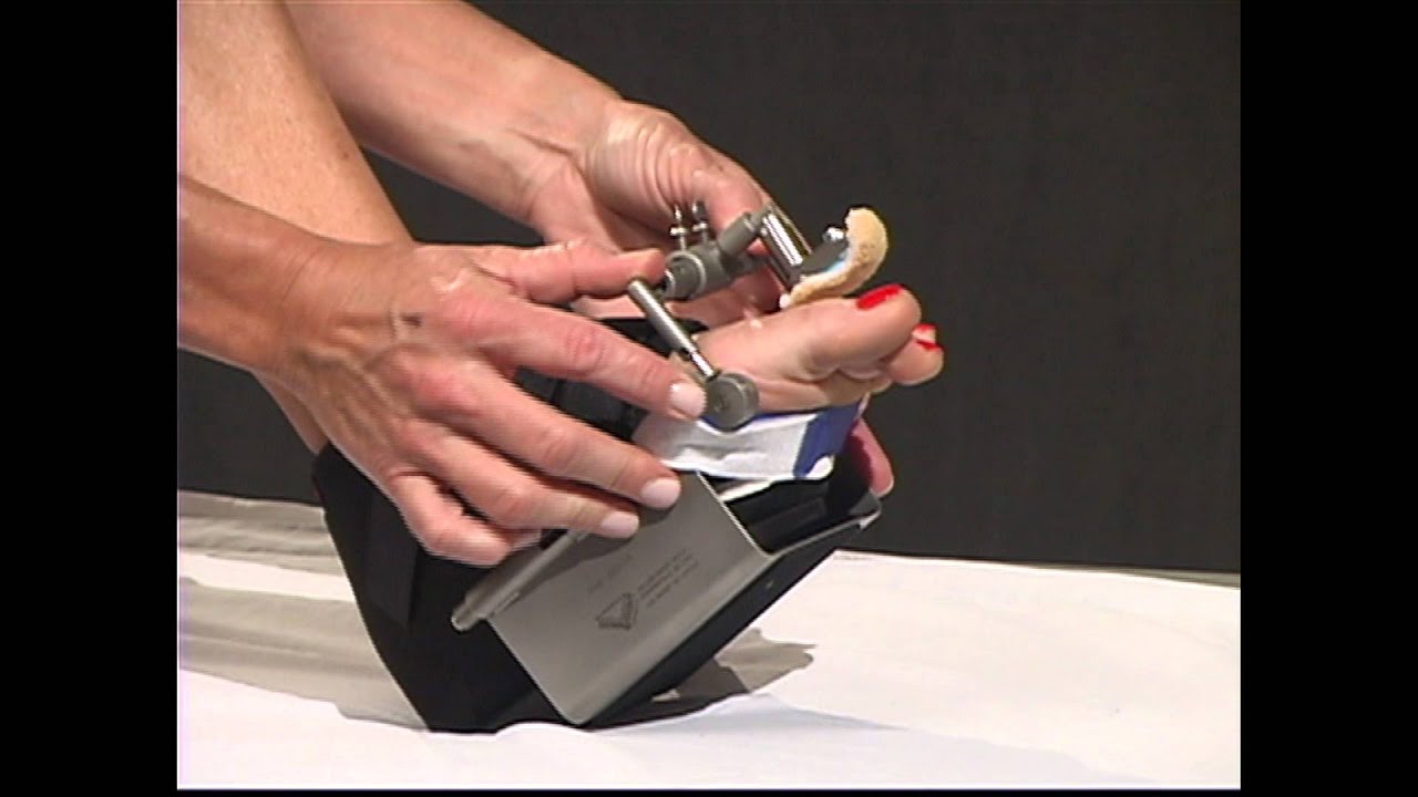 Symptoms SymptomsHammertoe and mallet toe feature an abnormal bend in the joints of one or more of your toes. Moving the affected toe may be difficult or painful. Corns and calluses can result from the toe rubbing against the inside of your shoes. See your doctor if you have persistent foot pain that affects your ability to walk properly. Diagnosis Your healthcare provider will examine your foot, checking for redness, swelling, corns, and calluses. Your provider will also measure the flexibility of your toes and test how much feeling you have in your toes. You may have blood tests to check for arthritis, diabetes, and infection. Non Surgical Treatment Prescription strength medicines to decrease pain and inflammation. Physical Therapy. To strengthen poorly functioning muscles and stretch tight muscles that may hammertoes be exacerbating the toes. Special ultrasound techniques may reduce inflammation. Custom Foot Orthotics. An orthotic with an exact mold of your foot to better align and support the foot to ease current discomfort and prevent future progression. Toe Splints or Pads. Specific pads may prevent pressure and physical irritation in shoes. Toe splints and toe spacers physically realign the toes and can lessen pain and halt or stall hammer toe progression. Cortisone injections are strong anti-inflammatory agents to decrease pain, and swelling directly at the toe region. Injections only treat the symptoms, and in some cases used in caution (and sparingly) they can weaken supporting ligaments of the toe(s). Surgical Treatment If these non-invasive treatments don?t work, or if the joint is rigid, a doctor?s only recourse may be to perform surgery. During the surgery, the doctor makes an incision and cuts the tendon to release it or moves the tendon away from or around the joint. Sometimes part of the joint needs to be removed or the joint needs to be fused. Each surgery is different in terms of what is needed to treat the hammertoe. Normally after any foot surgery, patients use a surgical shoe for four to six weeks, but often the recovery from hammertoe surgery is more rapid than that. An unfortunate reality is that hammertoe can actually return even after surgery if a patient continues to make choices that will aggravate the situation. Though doctors usually explain pretty clearly what needs to be done to avoid this.  Prevention PreventionThe number-one hammertoe prevention tip is to wear properly fitting shoes. If your shoes feel too snug, go to your local shoe store and have the length and width of your feet measured. If you wear high heels, keep the heel height to 2 inches or less. Wearing shoes with high heels increases the pressure on your toes and causes them to bend. It can also cause the formation of corns and a high arch. 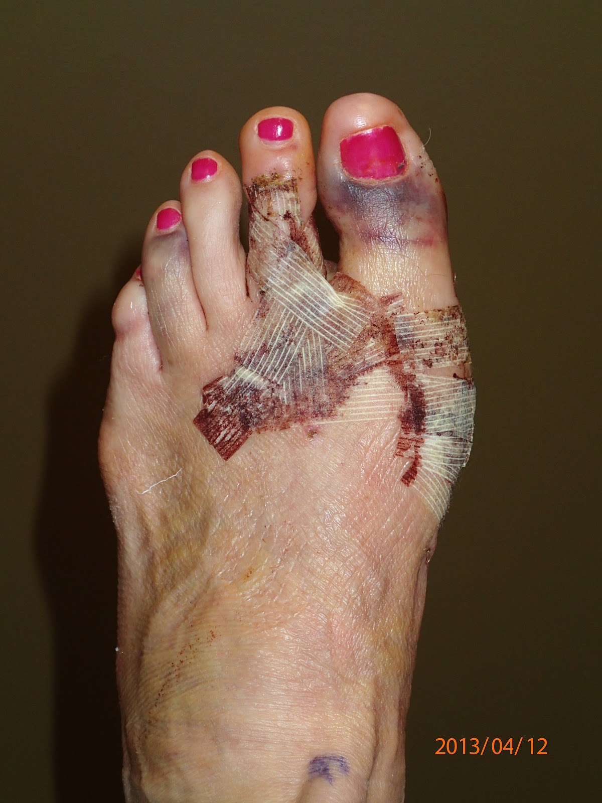 Overview OverviewA Hammer toes is a toe that is bent because of a weakened muscle. The weakened muscle makes the tendons (tissues that connect muscles to bone) shorter, causing the toes to curl under the feet. Hammertoes can run in families. They can also be caused by shoes that are too short. Hammertoes can cause problems with walking and can lead to other foot problems, such as blisters, calluses, and sores. Splinting and corrective footwear can help in treating hammertoes. In severe cases, surgery to straighten the toe may be necessary. Causes Wearing shoes that squeeze the toes or high heels that jam the toes into the front of the shoe. Other causes or factors in the development of hammertoes can include an injury such as badly stubbing your toe, arthritis and nerve and muscle damage from diseases such as diabetes. And, hammertoes tend to run in families, although it is more likely the faulty foot mechanics that lead to hammertoes that are inherited, not the hammertoes themselves. Hammertoe generally affect the smaller toes of the foot, especially the second toe, which for many people is the longest toe. It's uncommon for the big toe to be bent this way. 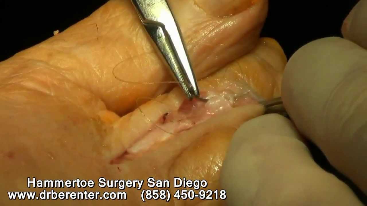 Symptoms SymptomsA hammertoe may be present but not always painful unless irritated by shoes. One may have enlarged toe joints with some thickened skin and no redness or swelling. However, if shoes create pressure on the joint, the pain will usually range from pinching and squeezing to sharp and burning. In long standing conditions, the dislocated joints can cause the pain of arthritis. Diagnosis Hammertoes are progressive, they don?t go away by themselves and usually they will get worse over time. However, not all cases are alike, some hammertoes progress more rapidly than others. Once your foot and ankle surgeon has evaluated your hammertoes, a treatment plan can be developed that is suited to hammertoes your needs. Non Surgical Treatment Conservative treatment is limited to accommodation, not correction, of the deformity, though some patients find the relief they can get from these options to be more than enough to put off or even avoid surgery. These include better Footwear. Shoe gear with a wider toe box and higher volume causes less friction to the toes. Toe Braces and Strapping. Some toe braces and strapping techniques take some pressure off the toes during gait. Custom molded orthotics can redistribute the forces through the tendons that control the toe, lessening the pain and extent of the deformity.The calluses on the toe and the ball of the foot can be shaved occasionally to reduce some pain and pressure, although they will return due to the constant deformity. Surgical Treatment In advanced cases in which the toe has become stiff and permanently bent, the toe can be straightened with surgery. One type of surgery involves removing a small section of the toe bone to allow the toe to lie flat. Surgery for hammertoe usually is classified as a cosmetic procedure. Cosmetic foot surgeries sometimes cause complications such as pain or numbness, so it?s better to treat the problem with a shoe that fits properly.
Overview
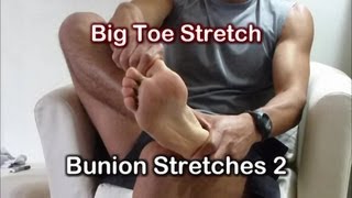 A bunion, or Hallux Valgus, is a foot deformity characterised by deviation of the bones around the big toe joint of the foot. As a result, there is a large exostosis or bony lump on the inside of the foot and the toe is pointed across towards the smaller toes. It is a common problem, more so in women and has been attributed to tight fitting footwear. This article will cover the basics of bunions, what it is due to, and what treatment is available. A bunion, or Hallux Valgus, is a foot deformity characterised by deviation of the bones around the big toe joint of the foot. As a result, there is a large exostosis or bony lump on the inside of the foot and the toe is pointed across towards the smaller toes. It is a common problem, more so in women and has been attributed to tight fitting footwear. This article will cover the basics of bunions, what it is due to, and what treatment is available.Causes Bunions are the byproduct of unnatural forces and motion being applied to the joints and tendons of your foot over a prolonged period of time. They can also be caused by traumas to the foot as well as congenital deformities. Occupations or athletic activities that place abnormal stress on your feet can also lead to the formation of bunions. Bunions have a tendency to run in families, but it?s not the bunion itself that is hereditary. It?s the the foot type which *causes* the bunion that is hereditary. Also, wearing shoes such as high heels that do not distribute your body weight evenly can lead to bunions, which explains why so many women suffer from bunions. Symptoms The most common complaint with bunions is pain that develops over the large bump due to shoe pressure. A red, inflamed area can develop, called a bursa. With ongoing pressure, the inflammation can cause throbbing, as it presses against the blood vessels, or swelling in the joint. Shooting pains occur when the swelling presses against the nerve. If left untreated, the constant irritation can lead to arthritis that breaks down the joint, resulting in pain and stiffness. Diagnosis Diagnosis begins with a careful history and physical examination by your doctor. This will usually include a discussion about shoe wear and the importance of shoes in the development and treatment of the condition. X-rays will probably be suggested. This allows your doctor to measure several important angles made by the bones of the feet to help determine the appropriate treatment. Non Surgical Treatment Separating the big toe and the next one with a 1 cm thick piece of foam can relieve painful symptoms. This can be fixed in place with some zinc oxide plaster tape and has the effect of straightening the great toe and relieving pressure on the bunion. A taping technique will support the joint and relieve the pressure on the inside of the foot. Off the shelf orthotic insoles can be worn to help correct any biomechanical problems in the foot which may be causing the problem. If the foot rolls in or over pronates then this causes the arch of the foot to flatten and more pressure is placed on the base of the big toe where the bunion forms. A podiatrist is a therapist who specializes in feet. They can do a full gait analysis and make orthotic inserts to correct biomechanical foot problems. Severe cases may require surgery to realign the joint but this is a last resort if conservative treatment has failed.  Surgical Treatment Most bunions can be treated without surgery. But when nonsurgical treatments are not enough, surgery can relieve your pain, correct any related foot deformity, and help you resume your normal activities. An orthopaedic surgeon can help you decide if surgery is the best option for you. Whether you've just begun exploring treatment for bunions or have already decided with your orthopaedic surgeon to have surgery, this booklet will help you understand more about this valuable procedure. Overview
Overpronation still continues to be misused and misunderstood. For example, there was a study that got a lot of recent mileage in the mainstream media and the blogosphere that claimed to show that foot pronation was not associated with injury risk. It was intriguing following comments on the study in mainstream media and in social media, especially the parroting of the press release without any critical appraisal. The study actually eliminated the ?overpronators? that were probably at high risk from the study then found that ?overpronation? was not a risk factor. What is more intriguing was that there was another study from around the same time that found the exact opposite. Clearly, the data on ?overpronation? and risk for injury in runners is mixed, so we need to rely on the more formal systematic reviews and meta-analyses of all the data. The most recent one of those concluded that ?overpronation? is just a small risk factor for running injury risk, but it is still statistically significant.  Causes In adults, the most common reason for the onset of Over-Pronation is a condition known as Post Tibial Tendonitis. This condition develops from repetitive stress on the main supporting tendon (Posterior Tibial Tendon) of the foot arch. As the body ages, ligaments and muscles can weaken. When this occurs the job of providing the majority of the support required by the foot arch is placed upon this tendon. Unfortunately, this tendon cannot bear the weight of this burden for too long. Eventually it fatigues under the added strain and in doing so the foot arch becomes progressively lower over a period of time. Symptoms Over-pronation is a condition where the arch flattens out which makes the feet roll inward while walking. This condition is also known as flat feet. It imposes extreme additional stresses on the plantar fascia, a fibrous band of tissue which connects the heel to the forefoot. Over-pronation makes walking a painful experience because of the additional strain on the calves, heel and/or back. Treatment for over-pronation involves the use of specially-made orthotics which offers arch support and medial rear foot posting as corrective measures. Diagnosis Your healthcare provider will ask about your symptoms, medical history, and activities and examine your feet. Your provider may watch you walk or run. Check the motion of your feet when they strike the ground. Look at your athletic shoes to see if they show an abnormal pattern of wear. 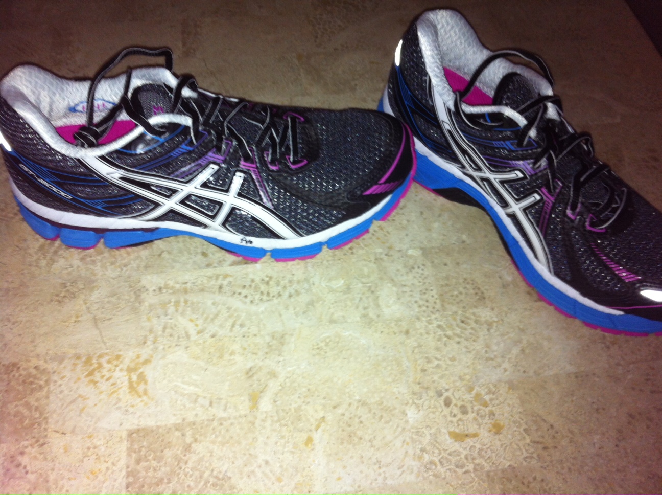 Non Surgical Treatment Supportive orthotics in the shoe is a method commonly implemented to treat many common running injuries associated with pronation. An advantage of orthotics is that they often allow the sufferer to continue to participate in athletic activity and avoid other treatment options that could be potentially costly and time consuming. Seventy-five percent of injured runners are successfully treated with the prescription of orthoses. Orthotics are the most effective treatment for symptoms that develop from unusual biomechanics within the body such as overpronation, resulting in either great improvement or complete healing of the injury in about half the cases. Surgical Treatment Subtalar Arthroereisis. The ankle and hindfoot bones/midfoot bones around the joint are fused, locking the bones in place and preventing all joint motion. This may also be done in combination with fusion at other joints. This is a very aggressive option usually reserved for extreme cases where no joint flexibility is present and/or the patient has severe arthritic changes in the joint. Overview
Sever's disease is a pain that occurs in the back of the heel of physically active children at around the time of puberty. Sever's disease occurs due to an inflammation of the growth plate due to excessive pull of the achilles tendon on this growth plate. During puberty the bones quite often grow faster than the muscles and tendons causing the tendons to become tight, this tightness then results in excessive pull on the back of the heel resulting in this painful condition. Causes During the growth spurt of early puberty, the heel bone (also called the calcaneus) sometimes grows faster than the leg muscles and tendons. This can cause the muscles and tendons to become very tight and overstretched, making the heel less flexible and putting pressure on the growth plate. The Achilles tendon (also called the heel cord) is the strongest tendon that attaches to the growth plate in the heel. Over time, repeated stress (force or pressure) on the already tight Achilles tendon damages the growth plate, causing the swelling, tenderness, and pain of Sever's disease. Such stress commonly results from physical activities and sports that involve running and jumping, especially those that take place on hard surfaces, such as track, basketball, soccer, and gymnastics. Symptoms The symptoms of Sever?s disease occur in the heel and the foot, and may worsen with activity. Pain and stiffness can occur in one or both heels. Symptoms can include. Swelling in the heel. Redness in the heel. Antalgic gait (such as limping). Foot pain or stiffness first thing in the morning or while walking. Pain that is worsened by squeezing the heel. Diagnosis A Podiatrist can easily evaluate your child?s feet, to identify if a problem exists. Through testing the muscular flexibility. If there is a problem, a treatment plan can be create to address the issue. At the initial treatment to control movement or to support the area we may use temporary padding and strapping and depending on how successful the treatment is, a long-term treatment plan will be arranged. This long-term treatment plan may or may not involve heel raises, foot supports, muscle strengthening and or stretching. Non Surgical Treatment Treatment is initially focused on reducing the present pain and limitations and then on preventing recurrence. Limitation of activity (especially running and jumping) usually is necessary. In Micheli and Ireland's study, 84% of 85 patients were able to resume sports activities after 2 months. If the symptoms are not severe enough to warrant limiting sports activities or if the patient and parents are unwilling to miss a critical portion of the sport season, wearing a half-inch inner-shoe heel lift (at all times during ambulation), a monitored stretching program, presport and postsport icing, and judicious use of anti-inflammatory agents normally reduce the symptoms and allow continued participation. If symptoms worsen, activity modification must be included. For severe cases, short-term (2-3 weeks) cast treatment in mild equinus can be used. Prevention Sever's Disease may be prevented by maintaining good flexibility while the child is growing. The stretching exercises can help lower the risk for injuries during a growth spurt. Having good quality shoes with firm support and a shock-absorbent sole will also help. Child should also avoid excessive running on hard surfaces.
Overview
Originally known as posterior tibial tendon dysfunction or insufficiency, adult-acquired flatfoot deformity encompasses a wide range of deformities. These deformities vary in location, severity, and rate of progression. Establishing a diagnosis as early as possible is one of the most important factors in treatment. Prompt early, aggressive nonsurgical management is important. A patient in whom such treatment fails should strongly consider surgical correction to avoid worsening of the deformity. In all four stages of deformity, the goal of surgery is to achieve proper alignment and maintain as much flexibility as possible in the foot and ankle complex. However, controversy remains as to how to manage flexible deformities, especially those that are severe.  Causes Posterior tibial tendon dysfunction is the most common cause of acquired adult flatfoot deformity. There is often no specific event that starts the problem, such as a sudden tendon injury. More commonly, the tendon becomes injured from cumulative wear and tear. Posterior tibial tendon dysfunction occurs more commonly in patients who already have a flat foot for other reasons. As the arch flattens, more stress is placed on the posterior tibial tendon and also on the ligaments on the inside of the foot and ankle. The result is a progressive disorder. Symptoms Posterior tibial tendon insufficiency is divided into stages by most foot and ankle specialists. In stage I, there is pain along the posterior tibial tendon without deformity or collapse of the arch. The patient has the somewhat flat or normal-appearing foot they have always had. In stage II, deformity from the condition has started to occur, resulting in some collapse of the arch, which may or may not be noticeable. The patient may feel it as a weakness in the arch. Many patients initially present in stage II, as the ligament failure can occur at the same time as the tendon failure and therefore deformity can already be occurring as the tendon is becoming symptomatic. In stage III, the deformity has progressed to the extent where the foot becomes fixed (rigid) in its deformed position. Finally, in stage IV, deformity occurs at the ankle in addition to the deformity in the foot. Diagnosis Your podiatrist is very familiar with tendons that have just about had enough, and will likely be able to diagnose this condition by performing a physical exam of your foot. He or she will probably examine the area visually and by feel, will inquire about your medical history (including past pain or injuries), and may also observe your feet as you walk. You may also be asked to attempt standing on your toes. This may be done by having you lift your ?good? foot (the one without the complaining tendon) off the ground, standing only on your problem foot. (You may be instructed to place your hands against the wall to help with balance.) Then, your podiatrist will ask you to try to go up on your toes on the bad foot. If you have difficulty doing so, it may indicate a problem with your posterior tibial tendon. Some imaging technology may be used to diagnose this condition, although it?s more likely the doctor will rely primarily on a physical exam. However, he or she may order scans such as an MRI or CT scan to look at your foot?s interior, and X-rays might also be helpful in a diagnosis. Non surgical Treatment The adult acquired flatfoot is best treated early. Accurate assessment by your doctor will determine which treatment is suitable for you. Reduce your level of activity and follow the RICE regime. R - rest as often as you are able. Refrain from activity that will worsen your condition, such as sports and walking. I - ice, apply to the affected area, ensure you protect the area from frostbite by applying a towel over the foot before using the ice pack. C - compression, a Tubigrip or elasticated support bandage may be applied to relieve symptoms and ease pain and discomfort. E - elevate the affected foot to reduce painful swelling. You will be prescribed pain relief in the form of non-steroidal antiinflammatory medications (if you do not suffer with allergies or are asthmatic). Immobilisation of your affected foot - this will involve you having a below the knee cast for four to eight weeks. In certain circumstances it is possible for you to have a removable boot instead of a cast. A member of the foot and ankle team will advise as to whether this option is suitable for you. Footwear is important - it is advisable to wear flat sturdy lace-up shoes, for example, trainers or boots. This will not only support your foot, but will also accommodate orthoses (shoe inserts).  Surgical Treatment If initial conservative therapy of posterior tibial tendon insufficiency fails, surgical treatment is considered. Operative treatment of stage 1 disease involves release of the tendon sheath, tenosynovectomy, debridement of the tendon with excision of flap tears, and repair of longitudinal tears. A short-leg walking cast is worn for 3 weeks postoperatively. Teasdall and Johnson reported complete relief of pain in 74% of 14 patients undergoing this treatment regimen for stage 1 disease. Surgical debridement of tenosynovitis in early stages is believed to possibly prevent progression of disease to later stages of dysfunction.
Overview Adult acquired flatfoot deformity (AAFD) is a painful condition resulting from the collapse of the longitudinal (lengthwise) arch of the foot. As the name suggests, this condition is not present at birth or during childhood. It occurs after the skeleton is fully matured. In the past it was referred to a posterior tibial tendon dysfunction (or insufficiency). But the name was changed because the condition really describes a wide range of flatfoot deformities. AAFD is most often seen in women between the ages of 40 and 60. This guide will help you understand how the problem develops, how doctors diagnose the condition, what treatment options are available.
 Causes Several risk factors are associated with PTT dysfunction, including high blood pressure, obesity, diabetes, previous ankle surgery or trauma and exposure to steroids. A person who suspects that they are suffering from PTT dysfunction should seek medical attention earlier rather than later. It is much easier to treat early and avoid a collapsed arch than it is to repair one. When the pain first happens and there is no significant flatfoot deformity, initial treatments include rest, oral anti-inflammatory medications and, depending on the severity, a special boot or brace. Symptoms Many patients with this condition have no pain or symptoms. When problems do arise, the good news is that acquired flatfoot treatment is often very effective. Initially, it will be important to rest and avoid activities that worsen the pain. Diagnosis The adult acquired flatfoot, secondary to posterior tibial tendon dysfunction, is diagnosed in a number of ways with no single test proven to be totally reliable. The most accurate diagnosis is made by a skilled clinician utilizing observation and hands on evaluation of the foot and ankle. Observation of the foot in a walking examination is most reliable. The affected foot appears more pronated and deformed compared to the unaffected foot. Muscle testing will show a strength deficit. An easy test to perform in the office is the single foot raise. A patient is asked to step with full body weight on the symptomatic foot, keeping the unaffected foot off the ground. The patient is then instructed to "raise up on the tip toes" of the affected foot. If the posterior tibial tendon has been attenuated or ruptured, the patient will be unable to lift the heel off the floor and rise onto the toes. In less severe cases, the patient will be able to rise on the toes, but the heel will not be noted to invert as it normally does when we rise onto the toes. X-rays can be helpful but are not diagnostic of the adult acquired flatfoot. Both feet - the symptomatic and asymptomatic - will demonstrate a flatfoot deformity on x-ray. Careful observation may show a greater severity of deformity on the affected side. Non surgical Treatment Although AAF is not reversible without surgery, appropriate treatment should address the patient?s current symptoms, attempt to reduce pain, and allow continued ambulation. In the early stages, orthotic and pedorthic solutions can address the loss of integrity of the foot?s support structures, potentially inhibiting further destruction.3-5 As a general principle, orthotic devices should only block or limit painful or destructive motion without reducing or restricting normal motion or muscle function. Consequently, the treatment must match the stage of the deformity. Causes Several risk factors are associated with PTT dysfunction, including high blood pressure, obesity, diabetes, previous ankle surgery or trauma and exposure to steroids. A person who suspects that they are suffering from PTT dysfunction should seek medical attention earlier rather than later. It is much easier to treat early and avoid a collapsed arch than it is to repair one. When the pain first happens and there is no significant flatfoot deformity, initial treatments include rest, oral anti-inflammatory medications and, depending on the severity, a special boot or brace. Symptoms Many patients with this condition have no pain or symptoms. When problems do arise, the good news is that acquired flatfoot treatment is often very effective. Initially, it will be important to rest and avoid activities that worsen the pain. Diagnosis The adult acquired flatfoot, secondary to posterior tibial tendon dysfunction, is diagnosed in a number of ways with no single test proven to be totally reliable. The most accurate diagnosis is made by a skilled clinician utilizing observation and hands on evaluation of the foot and ankle. Observation of the foot in a walking examination is most reliable. The affected foot appears more pronated and deformed compared to the unaffected foot. Muscle testing will show a strength deficit. An easy test to perform in the office is the single foot raise. A patient is asked to step with full body weight on the symptomatic foot, keeping the unaffected foot off the ground. The patient is then instructed to "raise up on the tip toes" of the affected foot. If the posterior tibial tendon has been attenuated or ruptured, the patient will be unable to lift the heel off the floor and rise onto the toes. In less severe cases, the patient will be able to rise on the toes, but the heel will not be noted to invert as it normally does when we rise onto the toes. X-rays can be helpful but are not diagnostic of the adult acquired flatfoot. Both feet - the symptomatic and asymptomatic - will demonstrate a flatfoot deformity on x-ray. Careful observation may show a greater severity of deformity on the affected side. Non surgical Treatment Although AAF is not reversible without surgery, appropriate treatment should address the patient?s current symptoms, attempt to reduce pain, and allow continued ambulation. In the early stages, orthotic and pedorthic solutions can address the loss of integrity of the foot?s support structures, potentially inhibiting further destruction.3-5 As a general principle, orthotic devices should only block or limit painful or destructive motion without reducing or restricting normal motion or muscle function. Consequently, the treatment must match the stage of the deformity.  Surgical Treatment Surgery should only be done if the pain does not get better after a few months of conservative treatment. The type of surgery depends on the stage of the PTTD disease. It it also dictated by where tendonitis is located and how much the tendon is damaged. Surgical reconstruction can be extremely complex. Some of the common surgeries include. Tenosynovectomy, removing the inflamed tendon sheath around the PTT. Tendon Transfer, to augment the function of the diseased posterior tibial tendon with a neighbouring tendon. Calcaneo-osteotomy, sometimes the heel bone needs to be corrected to get a better heel bone alignment. Fusion of the Joints, if osteoarthritis of the foot has set in, fusion of the joints may be necessary. Surgical Treatment Surgery should only be done if the pain does not get better after a few months of conservative treatment. The type of surgery depends on the stage of the PTTD disease. It it also dictated by where tendonitis is located and how much the tendon is damaged. Surgical reconstruction can be extremely complex. Some of the common surgeries include. Tenosynovectomy, removing the inflamed tendon sheath around the PTT. Tendon Transfer, to augment the function of the diseased posterior tibial tendon with a neighbouring tendon. Calcaneo-osteotomy, sometimes the heel bone needs to be corrected to get a better heel bone alignment. Fusion of the Joints, if osteoarthritis of the foot has set in, fusion of the joints may be necessary.
|
